
Laser Pilonidal Sinus Treatment: Recovery Without Weeks of Dressings
The traditional operation for pilonidal sinus meant an open wound and two to three months of daily dressings. Laser and endoscopic treatment changed that entirely — here is what recovery actually looks like.
For decades, the standard treatment for a pilonidal sinus was wide excision with the wound left open to heal by secondary intention. It cured the disease reliably. It also left a large cavity in the natal cleft that took eight to twelve weeks to fill in, requiring daily dressings throughout, with all the time off work that implies.
For a condition that mostly affects people in their twenties and thirties who are working or studying, that was a substantial cost. Minimally invasive techniques address it directly.
How laser treatment works
Laser closure, sometimes called SiLaC, follows a simple logic. The problem is a cavity lined with unhealthy tissue and containing hair. If you clear the contents and destroy the lining, the cavity has nothing keeping it open and collapses.
The pits are excised through incisions of a few millimetres. The cavity is thoroughly curetted — every hair and all granulation tissue removed, since anything left behind will keep the process going. A radial laser fibre is then introduced and withdrawn slowly, delivering energy circumferentially to ablate the lining and shrink the cavity walls.
What is left is a few small openings rather than an open trench.
EPSiT: adding direct vision
Endoscopic pilonidal sinus treatment uses a fine endoscope passed into the cavity, so the surgeon can see inside it while working.
The advantage is thoroughness. Every branch and recess is visualised and cleared under direct vision, rather than cleaned by feel. For complex sinuses with multiple tracks — a common reason for recurrence — this matters considerably.
The two techniques are often combined: the endoscope to find and clear, the laser to seal.
The day of surgery
- Hair in the area is clipped beforehand
- Spinal or general anaesthesia; in limited disease, local anaesthesia with sedation is possible
- The procedure takes twenty to sixty minutes depending on the extent
- All pits and tracks are identified, sometimes with the help of dye
- The cavity is cleared completely, then ablated with laser or endoscopically
- A light dressing only — no packing
- Home the same day in most cases
Recovery, week by week
- Days 0 to 2: mild to moderate soreness controlled with oral painkillers. Avoid sitting directly on the area; use a cushion. Keep the dressing dry
- Days 3 to 7: most desk-based patients return to work. Some serous discharge is expected and normal. Daily gentle cleaning as instructed
- Weeks 2 to 4: the small wounds close progressively. Sitting becomes comfortable. Driving usually resumes within one to two weeks
- Weeks 4 to 8: complete healing in most cases. Gym and heavier activity resumed once cleared
- From month 2: hair reduction started or continued, and maintained for at least a year
Practical tips for the first fortnight
- Use a soft cushion or sit slightly to one side rather than directly on the cleft
- Wear loose cotton underwear and avoid tight jeans
- Keep the area completely dry — pat dry thoroughly after showering, and use a hairdryer on cool if needed
- Avoid long drives and long uninterrupted periods of sitting
- Shower rather than bathe; avoid swimming pools until fully healed
- Do not apply powders or antiseptics unless specifically advised
What is normal and what is not
- Normal: clear or lightly blood-stained discharge for one to two weeks
- Normal: mild soreness on sitting for the first week or two
- Normal: the wound taking several weeks to close fully
- Call the clinic: increasing pain, swelling or redness after the first few days
- Call the clinic: thick purulent discharge or an offensive smell
- Call the clinic: fever
- Call the clinic: a wound that seems to close and then swells again — this may indicate a collection underneath
If it does not heal completely
A proportion of patients have a small track that persists after laser or endoscopic treatment. This is not unusual and it is not a disaster.
The procedure is repeatable, and because so little tissue has been removed, every other option remains open — a repeat laser treatment, a pit picking procedure, or if the disease turns out to be more extensive than expected, a flap procedure.
The trade-off is worth stating plainly: minimally invasive treatment accepts a somewhat higher chance of needing a second procedure in exchange for a dramatically easier recovery from the first. For most patients that is a good deal. For someone who has already had multiple recurrences, a definitive flap procedure may be the better first choice.
The part that determines whether it stays away
Laser treatment deals with the existing cavity. It does nothing to stop new hair entering the cleft, which is what caused the problem in the first place.
Sustained hair reduction from the natal cleft and surrounding area for at least a year is the single most effective thing you can do to prevent recurrence. Laser hair reduction is more effective than shaving; frequent shaving leaves sharp stubble that penetrates skin more easily.
Alongside that: keep the area dry, avoid uninterrupted sitting, and address weight where relevant. None of this is glamorous advice, but it is what separates patients who have one procedure from those who have three.
Need this assessed? Read the full treatment page for pilonidal sinus treatment, or book an appointment at Dynamic Surgical Hospital, Ahmedabad.
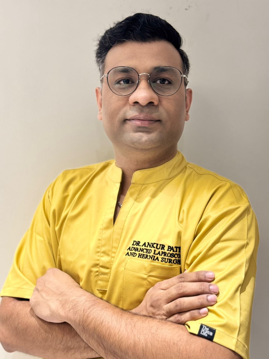
Dr. Ankur Patel
MBBS, MS (General Surgery), FMAS, FALS, DIPMAS, FACRSI
Dr. Ankur Patel is an advanced laparoscopic and robotic surgeon practising in Ahmedabad, with 15+ years of experience in minimally invasive and complex abdominal surgery. His work centres on hernia repair, gallbladder and appendix surgery, laser proctology and gastrointestinal procedures performed through keyhole techniques that shorten hospital stays and speed up recovery.
Read full profile →Medical disclaimer: This article is general patient education and is not a substitute for individual medical advice. Symptoms that seem similar can have very different causes, and only an examination can establish what is actually going on in your case. Please consult a qualified doctor and discuss your symptoms to better understand your condition and the right next steps for you. If you may be having a medical emergency, go to the nearest hospital.